Compact Bone Diagram Central Canal : bones tissue - Anatomy & Physiology 201 with Washo-krupps ... / (also, volkmann's canal) channel that branches off from the central canal and houses vessels and nerves that extend to the periosteum and endosteum.
Compact Bone Diagram Central Canal : bones tissue - Anatomy & Physiology 201 with Washo-krupps ... / (also, volkmann's canal) channel that branches off from the central canal and houses vessels and nerves that extend to the periosteum and endosteum.. Compact bone is very different from the other tissues you have seen. At the center of the osteon is a central canal we call the. A structural unit of compact bone consisting central haversian canal. These cylinder shaped structures are called osteons or haversian systems. So, you'll have these transverse canals that will lead into the central canal.
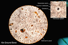
You can see, obviously, that blood supply has to move into the bone. Basically, in kindergarten when you drew skeletons, you were osteons are structural units of compact bone. Compact bone forms the surface of all bones. (b) in this micrograph of the osteon, you can clearly see the concentric lamellae and central canals. Its repeated pattern is arranged in concentric layers of solid bone tissue.
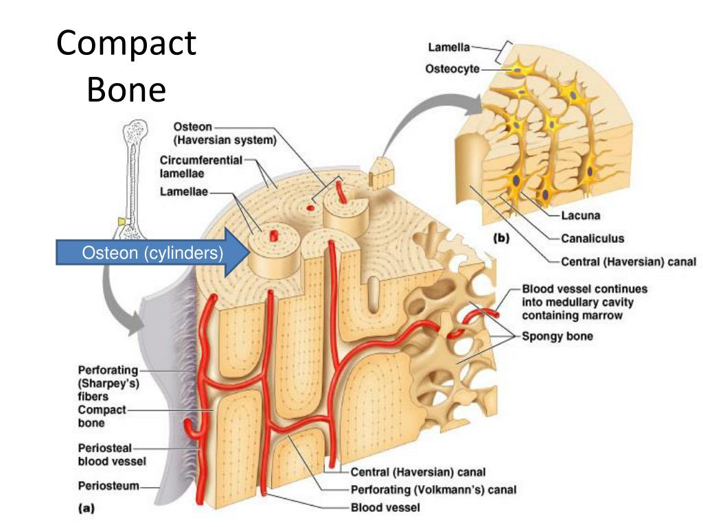
Osteon - Wikipedia from upload.wikimedia.org Compact bone consists of closely packed osteons or haversian systems. Long bone epiphyses ends of long bone thin layer of compact enclosing an area filled with spongy bone articular cartilage instead of periosteum, covers its external surface a glassy hyaline cartilage provides a smooth, slippery surface that decreases friction at joint surfaces long bone epiphyseal. (also, volkmann's canal) channel that branches off from the central canal and houses vessels and nerves that extend to the periosteum and endosteum. A structural unit of compact bone consisting central haversian canal. Mature compact bone is structurally layered or lamellar. In a compact bone, the haversian canals are tubes around narrow channels formed by lamellae. Compact bone is dense so that it can withstand compressive forces, while spongy (cancellous) bone has open spaces and supports shifts in weight an osteon comprises a long, hollow central canal that is surrounded by concentric layers called lamallae. Basically, in kindergarten when you drew skeletons, you were osteons are structural units of compact bone.
In the center of each osteon is the central canal, a space that houses blood vessels and.
Run perpendicular to the haversian canals and these connect osteons to one another and also as you can see carry their own set of small blood. The outlined area is a cross section of an osteon of compact bone. In the center of each osteon is the central canal, a space that houses blood vessels and. Between the rings of matrix, the bone cells (osteocytes) are located in spaces called lacunae. Edraw is a new uml diagram and software diagram drawing tool. Between the rings of matrix, the bone cells (osteocytes) are located in spaces called lacunae. In a compact bone, the haversian canals are tubes around narrow channels formed by lamellae. The osteon units of bone are made up of haversian canals (hc) and volkmann canals (vc), which run perpendicular. Compact bone is very different from the other tissues you have seen. Central canal compact bone perforating (vo. These cylinder shaped structures are called osteons or haversian systems. To recognise bone and understand its structure and to understand the processes by which bone can be formed. Compact bone is dense bone tissue found on the outside of a bone.
Each osteon consists of a central canal, which contains nerve filaments and one or two blood vessels, surrounded by lamellae. Compact bone consists of closely packed osteons or haversian systems. Compact bone also called cortical bone dense bone in which the bony matrix is solidly filled with organic ground substance and inorganic salts leaving only tiny spaces lacunae that contain the osteocytes or bone cells. The canal houses blood vessels, lymph vessels and nerves. Run perpendicular to the haversian canals and these connect osteons to one another and also as you can see carry their own set of small blood.
Chapter 6: Osseous Tissue and Bone Structure Flashcards ... from www.easynotecards.com In three dimensions an osteon is cylindrical in shape. Long bone epiphyses ends of long bone thin layer of compact enclosing an area filled with spongy bone articular cartilage instead of periosteum, covers its external surface a glassy hyaline cartilage provides a smooth, slippery surface that decreases friction at joint surfaces long bone epiphyseal. To recognise bone and understand its structure and to understand the processes by which bone can be formed. Central canal compact bone perforating (volkmann) al trabeculae periosteum osteon reset zoom. Osteons are long cylinders of bone that run parallel to the long axis of bone. Most bones contain compact and spongy osseous tissue, but their distribution and concentration vary based on the bone's overall function. Cancellous bones, compact bone, cortical bone, diaphyses, haversian canal, lamella, marrow cavity, osseous tissue, osteons, spongy bone this produces several concentric layers of lamellae around a central canal, forming an osteon. Compact bone forms the surface of all bones.
Compact bone consists of closely packed osteons or haversian systems.
The outlined area is a cross section of an osteon of compact bone. Compact bone is dense bone tissue found on the outside of a bone. Central (haversian) canal residences of osteocytes. The central region of compact bone consist of osteons. It is penetrated by a detailed system of haversian systems and interlinking vascular the cortical bone cells appear to be closely clustered together into a compact mass. In a compact bone, the haversian canals are tubes around narrow channels formed by lamellae. ( ) each osteon has a central haversian canal , running parallel to long axis of bone. The osteon consists of a central canal called the osteonic (haversian) canal, which is surrounded by concentric rings (lamellae) of matrix. Bone consists of cells along with the extracellular matrix produced by some of those cells. The canal houses blood vessels, lymph vessels and nerves. Run perpendicular to the haversian canals and these connect osteons to one another and also as you can see carry their own set of small blood. (also, volkmann's canal) channel that branches off from the central canal and houses vessels and nerves that extend to the periosteum and endosteum. Compact bone consists of closely packed osteons or haversian systems.
Microscopically compact bone has the features elucidated in the video (osteons), while the spongy bone is less dense and shows a framework of trabeculae. This extracellular matrix is composed of both organic and along these lamellae are spaces called lacunae, which contain cells called osteocytes. Long bone epiphyses ends of long bone thin layer of compact enclosing an area filled with spongy bone articular cartilage instead of periosteum, covers its external surface a glassy hyaline cartilage provides a smooth, slippery surface that decreases friction at joint surfaces long bone epiphyseal. Sclerostin inhibits bone formation mostly by antagonizing lrp5/6, thus inhibiting wnt signaling. Compact bone is made of concentric layers of osteocytes and bony matrix.
PPT - Long Bone Anatomy PowerPoint Presentation, free ... from image3.slideserve.com Sclerostin inhibits bone formation mostly by antagonizing lrp5/6, thus inhibiting wnt signaling. Central canal compact bone perforating (vo. Bone consists of cells along with the extracellular matrix produced by some of those cells. Compact bone, dense bone in which the bony matrix is solidly filled with organic ground substance and inorganic salts, leaving only tiny spaces that contain the osteocytes, or bone cells. A diagrammatic view of a cross section of bone. Central canal compact bone perforating (volkmann) al trabeculae periosteum osteon reset zoom. The outlined area is a cross section of an osteon of compact bone. Compact bone surrounds the spongy bone tissue and it has a unique appearance.
Between the rings of matrix, the bone cells (osteocytes) are located in spaces called lacunae.
Osteons are long cylinders of bone that run parallel to the long axis of bone. You can see, obviously, that blood supply has to move into the bone. Central canal compact bone perforating (volkmann) al trabeculae periosteum osteon reset zoom. Edraw is a new uml diagram and software diagram drawing tool. The osteon consists of a central canal called the osteonic (haversian) canal, which is surrounded by concentric rings (lamellae) of matrix. Central (haversian) canal residences of osteocytes. To recognise bone and understand its structure and to understand the processes by which bone can be formed. Microscopically compact bone has the features elucidated in the video (osteons), while the spongy bone is less dense and shows a framework of trabeculae. The compact bone is a dense bone found in the diaphysis. (also, volkmann's canal) channel that branches off from the central canal and houses vessels and nerves that extend to the periosteum and endosteum. These cylinder shaped structures are called osteons or haversian systems. This kind of bone tissue is not entirely solid even though they are. The outlined area is a cross section of an osteon of compact bone.
A structural unit of compact bone consisting central haversian canal compact bone diagram. Long bone epiphyses ends of long bone thin layer of compact enclosing an area filled with spongy bone articular cartilage instead of periosteum, covers its external surface a glassy hyaline cartilage provides a smooth, slippery surface that decreases friction at joint surfaces long bone epiphyseal.


0 Komentar